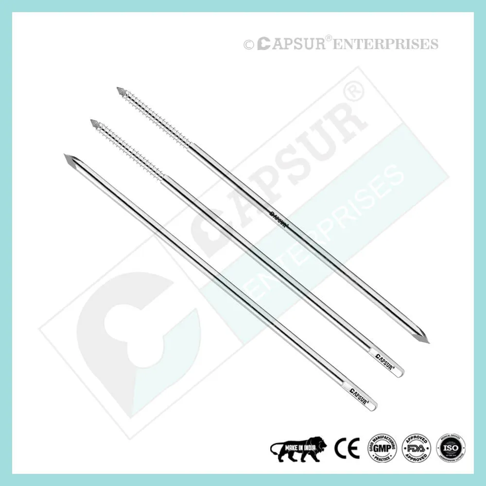
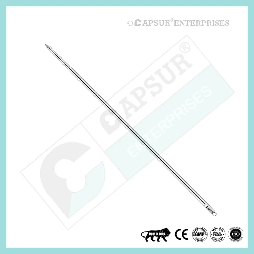
K Wire (Kirschner Wire) General principles
- The size of the K Wire (Kirschner Wire) is selected based on the child’s age and the size of the fragment.
- Choosing the right entry point and K-wire direction is essential for successful fixation.
- For the majority of simple fractures, two or three Kirschner wires (K-wires) provide adequate stabilization if the K-wires:
- the appropriate size (1.6/2.0 mm)
- Avoid rubbing shoulders at the fracture level.
- K Wire (Kirschner Wire) osteosynthesis placed intraosseously frequently necessitates additional plaster cast protection.
Advantages:
- Cheap
- widely accessible
- can be inserted manually (using a T-handle) or with a drill as long as thermal injury is prevented
- Simple to remove Drawbacks:
- Not consistently functional
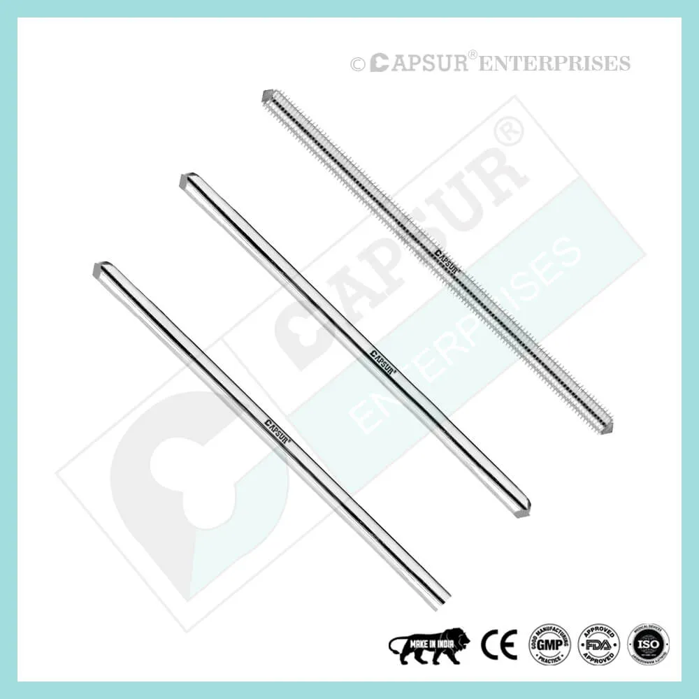
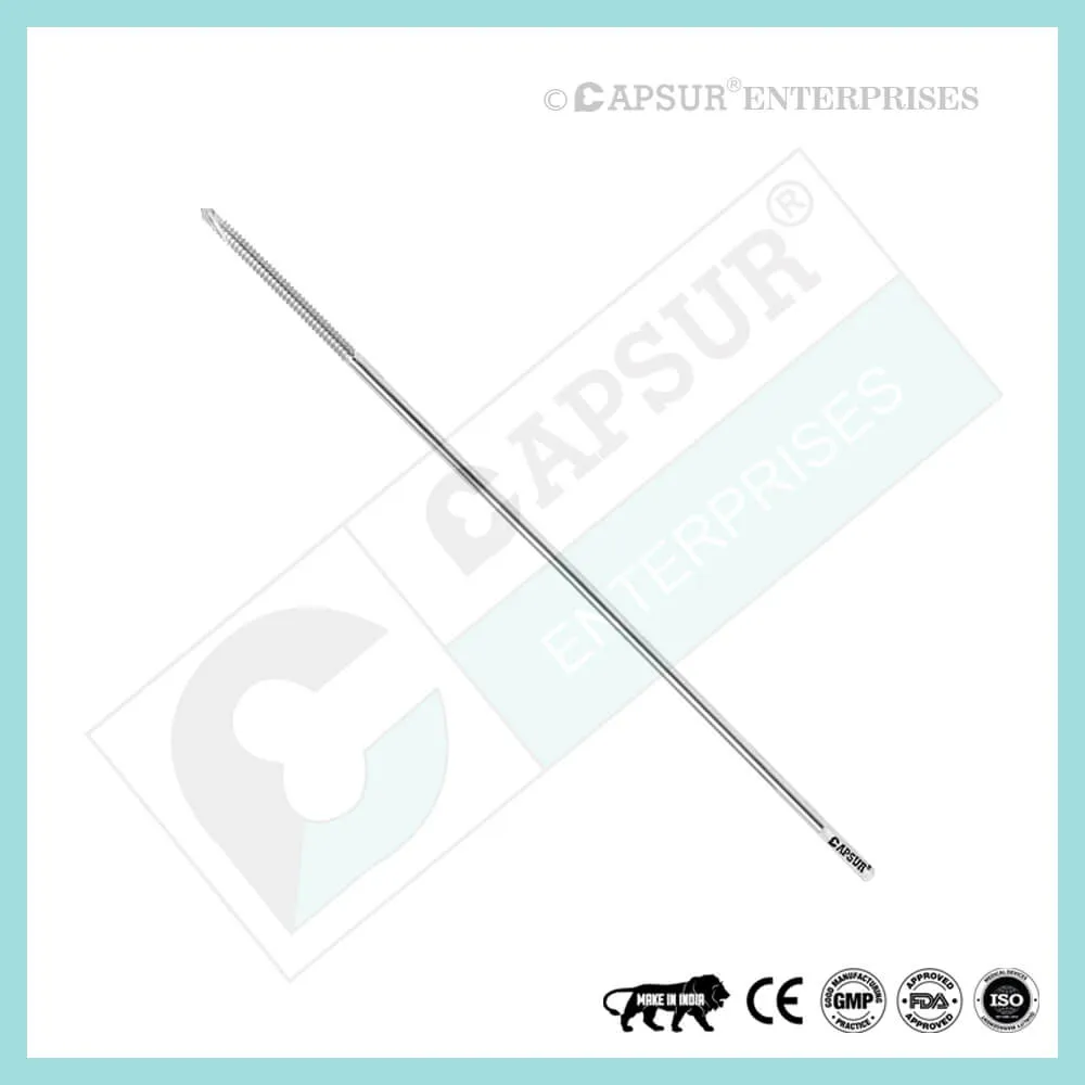
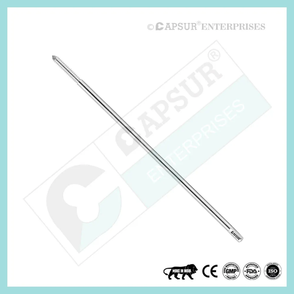
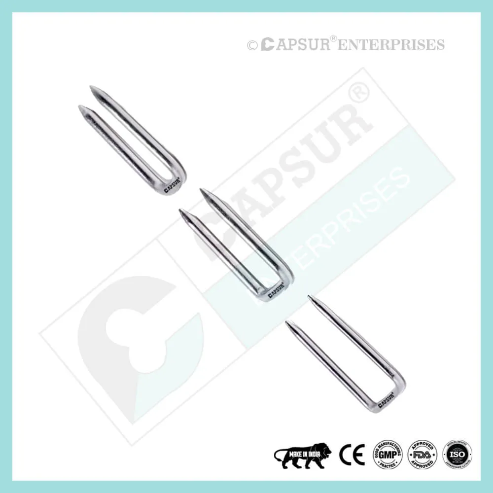
- Size of Kirschner Wire, or K Wire
- The K Wire’s (Kirschner Wire) size is affected by the factors listed below.
- Patient’s age and weight
- 1.6 mm K Wires (Kirschner Wires) are used for fractures near the shoulder, elbow, knee, and ankle joints in children younger than 5 to 6 years old.
- 2.0 mm K Wires (Kirschner Wires) are typically used in kids over this age.
- The patient’s weight must be taken into account when determining the K-wire’s diameter.
- Fracture site
- Long bone metaphyseal fractures require Kirschner Wires of at least 1.6 mm. Larger diameter wires might be required if only two wires are used.
- Small bone fractures (hand and foot) call for 1.0 to 1.6 mm K-wire.
sized fragments
The size of the fragment should be taken into consideration when selecting the K Wire (Kirschner Wire) size. For instance, a fracture of the lateral humeral condyle necessitates a K-wire with a smaller diameter than a fracture of the medial epicondyle of the humerus.
K Wire (Kirschner Wire) trajectory One size larger K-wires are used for bilaterally crossed K-wiring than for fractures fixed with two (or three) K Wires (Kirschner Wires) from only one side.
For instance, 1.6 mm K-wires can be used for bilateral crossed K-wiring of a supracondylar humeral fracture, whereas 2.0 mm K-wires are preferred for radial divergent wiring of the same fracture.
Kirschner Wire, or K Wire preparing the entry point for K Wire (Kirschner Wire)
In most instances, K Wires (also known as Kirschner Wires) are inserted from the free fragment into the main fragment. This makes it possible to control the free fragment using the K-wire as a joystick.
Where the K Wires (Kirschner Wires) cross the fracture line, the entry points should be placed as far apart as possible. This ensures the greatest possible rotational stability.
The entry point selection must be in accordance with the intended K-wire direction and the main fragment’s end fixation point.
If the anatomical site allows, the K Wires (Kirschner Wires) should ideally be inserted as perpendicularly to the fracture plane as possible. In some locations, this is not possible, so strict adherence to the aforementioned principle shouldn’t be allowed at the expense of mechanical stability.
Kirschner Wire (K Wire) transverse direction
It is important to choose the K Wires’ direction so that they are well separated at the fracture level.
To do this, the fracture line’s length is divided into four equally sized segments. When using two K-wires, the wires should ideally pass through the illustration’s green regions.
Kirschner Wires, a single-sided diverging pair of K wires
Fixation for oblique (>30°) metaphyseal fractures may be extremely challenging or impossible because at least one of the K Wires (Kirschner Wires) will be almost parallel to the fracture line.
Therefore, divergent monolateral K-wire fixation is preferable for oblique fractures. K Wires (Kirschner Wires) that are one size larger than those used for cross K-wiring should be used for this technique.
Another stabilization technique (such as an external fixator or plate) should be used if lateral divergent K Wire (Kirschner Wire) fixation is not possible, for instance because of soft tissue condition or a structure at risk.
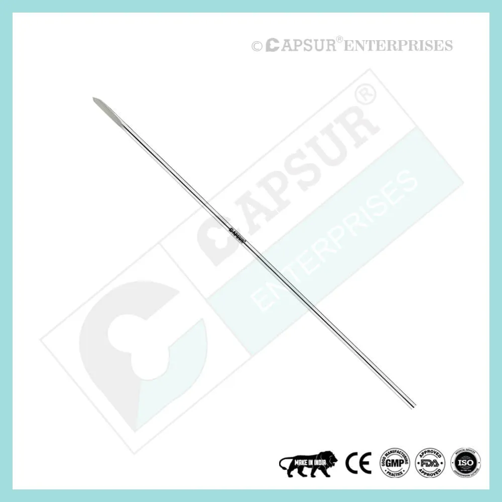
Stab incision with K Wire (Kirschner Wire) insertion
Over the intended entry point, a small incision or direct puncture with the K-wire is made. To prevent skin damage, which could result in a pin-track infection, an incision is advised.
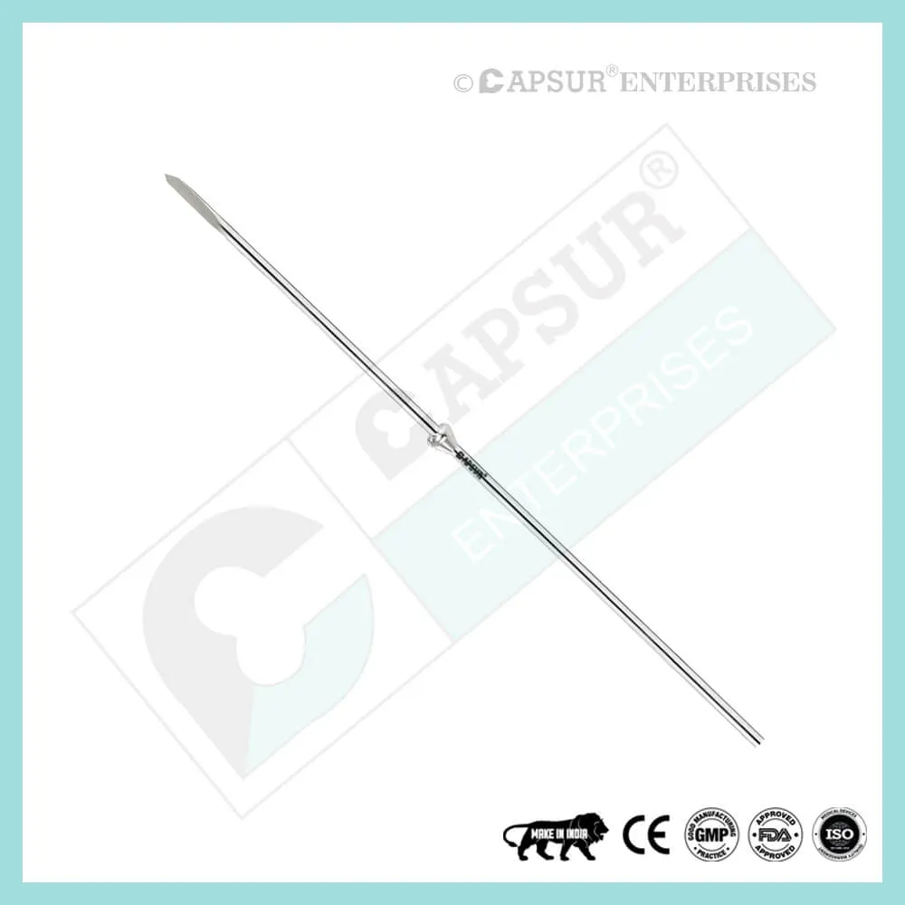
Insertion of the K Wire (Kirschner Wire)
K Wires (Kirschner Wires) should be inserted manually or with an oscillating drill to prevent thermal injury, particularly to the physis.
To prevent a thermal effect, a standard drill must be operated as slowly as possible.
Furthermore, while drilling, irrigate the K Wire (Kirschner Wire) with a cooled irrigation fluid.
The K Wire (Kirschner Wire) is initially manually inserted through the skin incision, onto the preferred bony entry point, if a drill is being used. The drill is fastened to the wire while holding the tip in its proper position.
The K-wire (Kirschner Wire) can be inserted using a suitable drill sleeve to stabilize the wire, protect the soft tissues, and ensure optimal direction, which can help prevent bending.
To prevent whipping of the wire and loss of trajectory, it is beneficial to shorten the Kirschner Wire (K Wire) sticking out of the drill.
The K Wire (Kirschner Wire) tip should initially be held as orthogonal to the bone surface as possible until the tip of the wire has a good purchase in order to prevent skidding.
The angulation of the K-wire should be corrected in accordance with the intended direction of the K-wire once the tip of the K Wire (Kirschner Wire) has attained a good purchase.
Check that the tip of the Kirschner Wire is inserted into the far cortex of the main fragment as soon as you feel more resistance.
The K-wire’s tip should extend no more than 2-3 mm into the far cortex, but it should do so only partially. This prevents soft tissue irritation and neurovascular harm.
The wire’s free end is typically left sticking out through the skin after being bent 180 degrees. The entrance wound around the wire is covered with a sterile dressing.
One wire should only be inserted across a physis once every two attempts. Multiple attempts to insert the wire can repeatedly puncture the physis, which can disrupt subsequent growth.
Removal of K Wire (Kirschner Wire)
Depending on the child’s age, the pattern of the injury, and any additional injuries, the treating surgeon will decide when to remove the K Wire (Kirschner Wire).
Depending on the child’s age, the fracture has healed to the point where redisplacement is extremely unlikely after 3–4 weeks, at which point the K-wires can be taken out.
K Wire (Kirschner Wire) Risk Factor
When determining the prognosis in each situation, contraindications—which may be partial or complete—must be taken into account. The following situations may necessitate the consideration of alternative management strategies:
either localized or systemic, acute or chronic infections.
Chronic inflammation may be local, systemic, or both.
serve as vascular, nervous, or muscular disease that puts the afflicted area in danger.
faulty bone structures that would prevent the implant from being adequately anchored.
All afflictions that may endanger the implant’s success and functionality.
Warnings and Precautionary for K Wire (Kirschner Wire)
The surgeon and support staff should read the safety instructions in this document as well as any product-specific information in the product description, surgical techniques, and/or brochures before using K Wire (Kirschner Wire).
Wire is designed, built, and produced with the utmost care using materials of the highest quality for medical applications. If used properly, these high-quality wire ensure the best working outcomes. As a result, the usage guidelines and safety advice below must be followed.
Inappropriate use of wire can result in injury to the operator, patients, or other people as well as tissue damage, premature wear and tear, instrument destruction, and instrument destruction.
The operating surgeon must actively participate in the medical care of their patients. The surgeon must have a complete understanding of the instruments, their limitations, and the surgical procedure. The surgeon and the surgical team are responsible for exercising caution in the selection and use of surgical instruments. Before using implants, adequate surgical training should be obtained.
Factors that could harm the operation’s success include:
- allergies to materials implanted.
- regional bone tumors.
- osteomalacia or osteoporosis.
- metabolic disturbances and systemic disease.
- drug and alcohol abuse.
- Excessive shock-producing physical activity that exposes the implant to blows and/or heavy loads.
- Patients who lack the mental capacity to comprehend and follow instructions from a doctor.
- Unhealthy overall.
- Potential Negative Effects
- The most frequent side effects following implantation are as follows:
- cyclic loading of the fixation site and/or tissue reaction to the implant may cause the wire to loosen.
- the two stages of infection.
- additional bone fracture brought on by abnormal stress or weakened bone structure.
- a hematoma or pressure-related pressure that causes temporary or permanent neural damage.
- Hematomas from wounds and slow wound healing.
- Venous thrombosis, pulmonary embolism, and cardiac arrest are examples of vascular disease.
- heterotopically ossifying.
- Due to the K Wire’s (Kirschner Wire’s) presence, there is pain and discomfort.
- Implant mechanical failure, such as bending, loosening, or breakage.
- Implant migration leading to injury.
Preoperative Planning for K Wire (Kirschner Wire)
Following a thorough clinical evaluation of the patient, the operation is planned. X-rays are also necessary to provide a clear picture of the bony anatomy and any associated deformities. A full size of K Wire (Kirschner Wire) and the appropriate implantation tools must be on hand at the time of the procedure.
The potential risks and complications related to the use of implants should be discussed with the patient by the clinician. If the patient has allergies to any of the implant materials, it is crucial to know this before surgery. Additionally, the patient needs to be made aware that the device’s performance cannot be guaranteed because problems may reduce its lifespan.
K Wire (Kirschner Wire) Precautions
- During reprocessing, verify that the instruments are functional and look for wear. Before using, replace any worn-out or broken instruments.
- It is advised to use the tools designated for this wire.
- Use caution when handling equipment, and put used bone-cutting tools in a sharps container.
- Always use suction and irrigation to remove any debris that may be produced during implantation or removal.
K Wire (Kirschner Wire) Warnings
When put through excessive force while being used, Kirschner wire, also known as K Wire, can break. We advise that the broken part be removed whenever possible and practical for the particular patient, though the surgeon will ultimately decide whether to do so based on the risk involved. Be aware that implants lack the natural bone’s strength. Significant loads may cause implants to fail.
The user’s glove or skin may be pinched or torn by the sharp edges or moving joints of some instruments, screws, and cut plates.
Be sure to get rid of any fragments that weren’t fixed during surgery.
While the surgeon will ultimately decide whether to remove the implant, we advise that fixation devices be taken out as soon as it is safe and practical for the specific patient and after their purpose as a healing aid has been fulfilled. To prevent refracture, wire removal should be followed by adequate post-operative management.
K Wire (Kirschner Wire) General Adverse Events
There are risks, side effects, and adverse events associated with all major surgical procedures. While there are many possible reactions, the following are some of the most frequent ones: issues related to anesthesia and patient positioning (such as nausea, vomiting, dental injuries, neurological impairments, etc.), thrombosis, embolism, infection, damage to nerve and/or tooth roots or other critical structures, such as blood vessels, excessive bleeding, damage to soft tissues, including swelling, abnormal scar formation, functional impairment of the musculoskeletal system, and pain.